Journal of Scientific Exploration, Vol. 18, No. 2, pp. 253-273, 2004 0892-3310/04
Electrodermal Presentiments of Future Emotions
DEAN I. RADIN
Institute of Noetic Sciences San Antonion Rd.
Petaluna CA 94952
e-mail: deanradin@noetic.org
.
Abstract _ In previously reported double-blind experiments, electrodermal activity (EDA) monitored during display of randomly selected photographs showed that EDA was higher before emotional photos than before calm photos (p = 0.002). This differential effect, suggestive of precognition, was dubbed « presentiment. » Three new double-blind experiments were conducted in an attempt to replicate the original studies using the same basic design, but with new physiological monitoring hardware, software, stimulus photos, subject populations, and testing environments.
The three replications involved 109 participants who together contributed 3,709 trials. The new studies again showed higher EDA before emotional photos than before calm photos = 0.001). All four experiments combined involved 133 participants and 4,569 trials; the associated weighted mean effect size (per trial) was e — 0.064 ± 0.015, over 4 standard errors from a null effect. As a more general test, presentiment predicts a positive correlation between pre-stimulus EDA and independently assessed emotionality ratings of the photo targets. The observed correlation across all four experiments was significantly positive (p = 0.008).
Consideration of alternative explanations, including expectation, sensory cues, hardware or software artifacts, inappropriate analyses, and anticipatory strategies, revealed no suitable candidates that could systematically generate the observed results. This series of four experiments, supported by successful replications conducted by other investigators, appears to demonstrate a small magnitude but statistically robust form of precognition in the human autonomic nervous system.
Keywords:
: electrodermal activity-precognition-autonomic nervous system anticipation
.
Introduction
Many people have experienced intuitive hunches or forebodings about future events that later turned out to be correct. Most such hunches can be attributed to unconscious inferences, others are undoubtedly coincidences, instances of selective memory, or due to forgotten expertise. However, sometimes a hunch seems so intrinsically unlikely and yet turns out to be valid, that one wonders whether such experiences, often on the edge of conscious awareness, might involve perception of future information. In a series of experiments designed to test this idea under double-blind conditions, I explored whether the human autonomic nervous system would be able to correctly anticipate exposure to randomly selected calm or emotional photographs (Radin, 1997).
Those initial studies provided evidence for what I called presentiment. I used this term, in contrast to precognition, as the latter implies conscious awareness (i.e., pre-cognition) of future events. Publication of the initial results in this journal prompted a number of other researchers to attempt to replicate the effect. Some of the replications focused primarily on electrodermal activity (EDA), as in the original studies (Bierman 2000; & Radin, 1997, 1998; Norfolk, 1999; Parkhomtchouck et 2002; Spottiswoode & May, 2003; Wildey, 2001). Others explored different physiological measurements, including functional magnetic resonance imaging (MRI; Bierman & Scholte, 2002) and heart rate variability (McCraty, 2002). All of the replications reported results consistent with the original findings.
This paper reviews the results of presentiment experiments that I conducted from 1996 through 2000, only the first of which was previously published (Radin, 1997) All these experiments were primarily proof-oriented rep-lications using different hardware and software implementations, subject populations, environmental conditions, and photo stimuli. Experiment 1 was the initial series of tests, Experiment 2 was a straightforward replication, Experiment 3 was conducted as a proof-of-principle demonstration in an industrial research laboratory, and Experiment 4 explored the use of a custom-designed psychophysiological monitoring device. To avoid repeating descriptions of common design elements, a brief overview of the general procedure will be described first and then appropriate details added as each experiment is discussed.
.
Method
Basic Experimental Procedure :
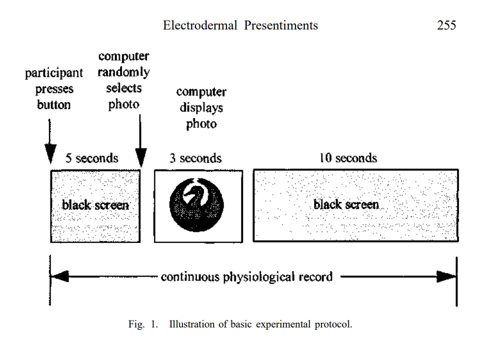
A participant (P) is seated in front of a computer monitor displaying a black screen. The experimenter attaches EDA electrodes (Ag-AgCl, 8 mm diameter) to the volar surfaces of the distal phalanges of the index and middle fingers of non-dominant hand. The electrodes are secured with Velcro straps, and electrode gel is used to enhance contact with the skin. After the experimenter ensures that the physiological hardware is recording the EDA data properly, P is instructed to press a button at will. When this occurs (see Figure 1), the computer waits 5 seconds, then it selects a photo at random from a large pool of possibilities, displays it for 3 seconds, and then the screen goes blank again for 10 seconds. After the 10-second « cool-down » period, the computer instructs P to press the button again when ready to begin the next trial. A typical session may last 30 minutes, during which time 40 trials are run. The design is double-blind in the sense that neither P nor the experimenter know in advance which photos will be displayed in a given session, or in what sequential order.
Hypotheses
The concept of presentiment postulates that present physiological states are correlated with near-term future experiences. When those futures involve emotional experiences, as evoked through the use of photographs of varying emotionality, the correlation is postulated to be detectable as a present-time arousal of the autonomic nervous system. This idea leads to three progressively more general hypotheses.
Hypothesis 1. This hypothesis states that EDA before display of emotional photos will be larger than EDA before display of calm photos. This differential prediction is tested by partitioning all trials in an experiment into emotional and calm subsets of equal size. These subsets are formed based upon prior assessments of the emotionality of the target photos. The probability of the resulting differences in EDA is formally tested using a nonparametric statistical method known as randomized permutation analysis (Blair & Karniski 1993).
Hypothesis 2. This hypothesis states that as the contrast between emotional and calm trials increases, the magnitude of the presentiment effect will also increase, and vice versa. This is tested by sorting all trials according to their pre- assessed emotionality ratings, then comparing the top 1 % most emotional vs. the 1% most calm trials, then the top 2%, and so on up to 50% (which is then the same as Hypothesis 1). No specific prediction is made for the emotional contrast level that would show the largest effect, but it is expected that the peak might fall somewhere between 5% and 25%. This is because a few photos with very high or very low emotionality assessments would provide strong emotional contrasts, but at the cost of low statistical power. And an assessment made with many photos of varying emotionality would provide greater statistical power, but at the cost of weaker emotional contrast.
Hypothesis 3. This hypothesis generalizes the first two hypotheses and predicts a positive correlation between the pre-assessed emotionality ratings and changes in EDA prior to the stimuli.
.
Method of Analysis
Electrodermal activity (EDA) refers to variations in electrical resistance of the skin. These fluctuations are due to activity of the eccrine sweat glands, which are activated by the sympathetic nervous system (Bouscein, 1992). The specific form of EDA used in these studies was skin conductance level (SCL). SCL has been used as the principal physiological measurement in these and in many of the replication attempts primarily because SCL is a conveniently measured and widely used indicator of overall autonomic activity. All SCL data in these experiments were uniformly analyzed using a simplification of the technique employed in the initial studies (Radin, 1997).
An SCL « sample » refers to an instantaneous measurement of absolute skin conductance. For the sake of exposition, let us assume in this discussion that one trial is 18 seconds in length (as shown in Figure 1) and that the sampling rate is 5 Hz; thus each trial consists of 90 samples. Let us further assume that an experiment consists of 20 participants, each of whom runs 30 trials, for a total of 600 trials.
To analyze the results of an experiment, each SCL sample in each trial is normalized as — where in our example refers to samples j= 1 to 600 trials, refers to the raw SCL value for sample in trial j, is the average of the 25 samples in the 5-second
period in j (i.e., from the starting button press to just before the photo appears), and is the standard deviation of those same 25 samples. Then, all normalized trials are clamped to zero after the button press as = —
where refers to the first normalized SCL sample after the button press in trial j, and ranges across all samples 1-90.
The values are thus changes in normalized SCL (i.e., ASCL). Normalized SCL is of interest, rather than absolute SCL, because otherwise a few Ps with highly labile SCL signals would overwhelm the data from other, less labile Ps. And change in SCL is of interest because we are interested in event-related responses, i.e., how autonomic arousal fluctuates from the moment P decides to begin each trial, rather than in general level of sympathetic arousal or in spontaneous fluctuations in SCL.
To determine the statistical likelihood of the differences in ASCL observed before emotional and calm trials, randomized permutation analysis was employed as follows (Blair & Karniski, 1993):